Patients need to understand! Which is your best choice: CT, DR, or MRI?
Patient 1: Hey, do you also do MRI scans here?
Patient 2: I have a headache! The doctor gave me a referral for a head MRI.
Family Member 3: My uncle suddenly had a headache! The doctor told him to get a head CT.
Doctor: Next, please. The patient for the head MRI, please follow me to get ready.
Patient 1: Hey! I'm here! Doctor, the two people sitting next to me also have headaches. Why are they getting a CT or a head MRI, but I have to get an enhanced head MRI with a needle?

The conversation above is something that patients often encounter when coming to the imaging department for an examination. Why is the examination different even though they all have headaches? In fact, many people may not clearly distinguish between different imaging tests. Today, I’ll briefly summarize the various imaging exams, their principles, advantages, and disadvantages to help you understand better.
DR Examination: Purpose and Applicable Scope
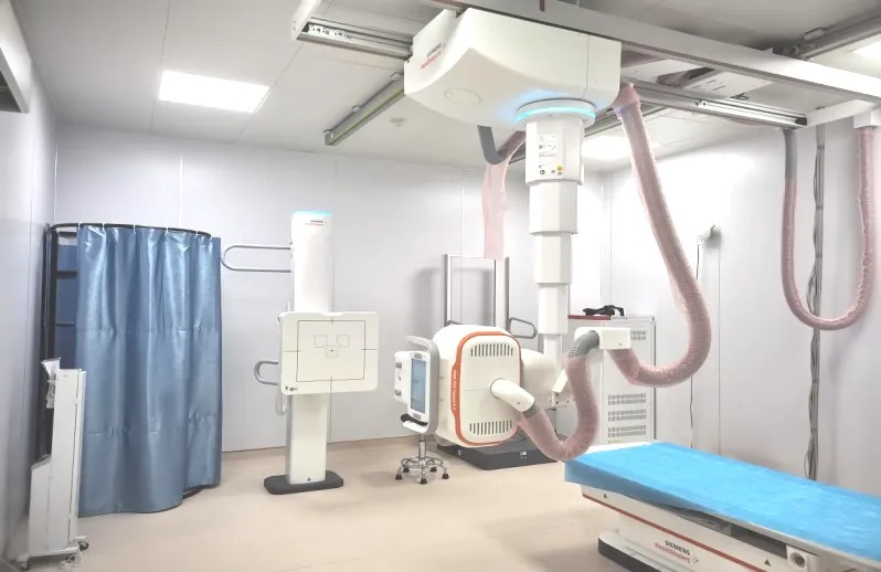
DR (Digital Radiography) uses X-rays to penetrate the human body and create black-and-white contrast images. It’s similar to traditional shadow puppetry, where the image is projected onto a screen to get overlapping images of the body’s structure (plain film). (Think of the X-ray tube as a flashlight, the detector as the screen, and the silhouette as the subject being examined.)
Advantages:
- Low radiation, affordable, and fast imaging.
- High spatial resolution.
Disadvantages:
- The overlapping structures in the plain film images may lead to interference, and soft tissue resolution is poor, which could result in missed or incorrect diagnoses. Its diagnostic value for solid organs and hidden fractures is limited.
- Not recommended for pregnant women or individuals highly sensitive to X-rays or those who should avoid exposure to X-rays.
Applicable Scope:
- Often used for initial screening or post-procedure checks (such as after catheter placement).
- Used for clinical examination in cases of acute abdominal conditions.
- The first choice for imaging in bone and joint diseases.

CT Examination: Purpose and Applicable Scope
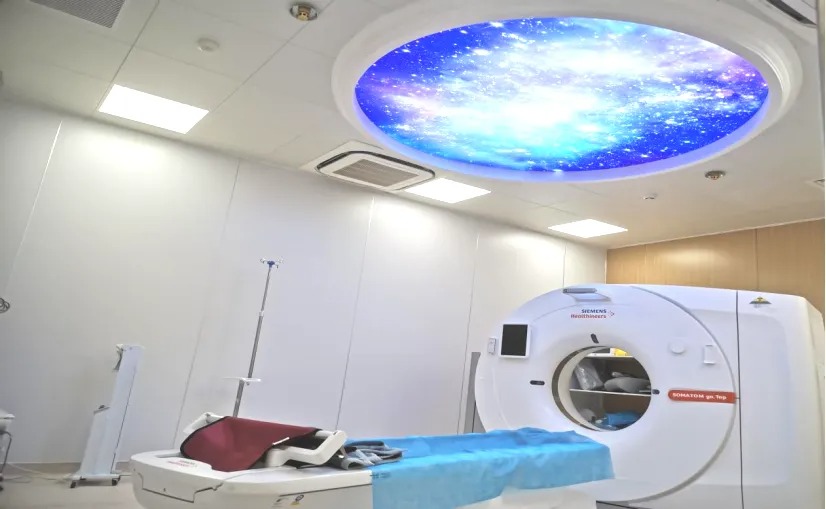
The imaging principle of CT (Computed Tomography) involves using X-ray beams to scan thin layers of the body at the examination site. It is similar to slicing a carrot into thin pieces, producing layer-by-layer images. (Think of the CT machine as the chef, the X-ray tube as the chef's knife, and the body as the carrot). Nowadays, spiral CT allows for continuous scanning, increasing the scanning speed and significantly reducing radiation exposure.
Advantages:
- Fast examination speed, easy operation, and fewer restrictions for the patient.
- High image density resolution.
- Clear scanning images, with easy differentiation of anatomical structures and their relationships.
- Provides cross-sectional images without overlapping tissues, and can be reconstructed in different planes (3D reconstruction).
- With the use of contrast agents, enhanced scans greatly improve the detection rate of lesions and assist with qualitative diagnosis and post-treatment evaluation.
Disadvantages:
- CT scanning exposes the patient to potential radiation hazards and has certain contraindications (e.g., pregnant women, individuals allergic to iodine contrast agents, etc.).
- Lower spatial resolution compared to other imaging methods.
- Lower soft tissue resolution compared to MRI, making it difficult to distinguish between tissues or lesions of similar densities.
Applicable Scope:
- CT is widely applicable for various purposes.
- Low-dose spiral CT can be used for chest exams in healthy individuals, early screening for lung nodules, or when necessary, CT scans for infants and young children.
- Conventional CT scans can be used for imaging various body parts, such as in cases of head trauma or acute, unexplained headaches, where a CT scan of the brain is used to rule out hemorrhages or large-scale brain ischemia.
- CT enhanced scans include routine enhancement (useful for detecting lesions in tumor patients and for follow-up monitoring), CT angiography, and CT perfusion imaging (which require intravenous contrast agents), and can be used for diagnosing and distinguishing diseases in various body systems, as well as for pre-operative or post-operative disease assessment.

MRI Examination: Purpose and Applicable Scope
Magnetic Resonance Imaging (MRI) is based on the principle that hydrogen protons in the human body align in an ordered manner when exposed to a magnetic field, similar to how iron filings align when they come into contact with a magnet. (The MRI magnet acts like a magnet, while the hydrogen protons in the body resemble the iron filings.)
Advantages:
- No ionizing radiation.
- Multi-axial and multi-parameter imaging.
- High soft tissue resolution with clearer anatomical structures, and the ability to perform various functional imaging (such as diffusion-weighted imaging, brain function imaging, etc.).
- MRI enhanced scans provide higher lesion detection rates and greater value for qualitative and quantitative disease analysis.
Disadvantages:
- Longer examination times, loud scanning noise, and higher requirements for patient preparation and cooperation during the scan.
- Several contraindications, such as patients with heart pacemakers, artificial metal heart valves, or other metal foreign objects; pregnant women in their first trimester; and patients with severe high fever (usually above 39°C) are not suitable for MRI scans.
Applicable Scope:
- MRI is widely applicable and can be used for imaging any part of the body.
- For older patients without a disease history, annual screening with a non-contrast head MRI and non-contrast head vascular scan should be a preferred choice.
- Routine MRI scans have a wide range of applications and can be used for initial screening of brain, spinal cord, neck, chest, breast, abdomen, pelvis, bone, joint, and muscle abnormalities.
- Enhanced MRI is of significant clinical value for follow-up assessments, efficacy evaluations, and tumor patients' monitoring.
Medical imaging plays a crucial role in modern clinical diagnostics and treatment. It provides intuitive and accurate information on human anatomy, helping clinicians to promptly detect diseases, assess conditions, formulate appropriate treatment plans, and follow up on the progression of diseases. It is also widely used in early disease screening. Therefore, whether it’s for early detection of lesions or monitoring treatment effectiveness, modern medical imaging plays an irreplaceable role.
For different diseases, selecting the most appropriate imaging method is essential for early diagnosis, which is what every patient and their family desires, and it is our goal as healthcare providers.


